- Home
- Image Gallery
Select an application or technique:
Latest Images
High-Speed Imaging
AFM / OT and Advanced Optics
Life Sciences
Polymers
Nanoscience
Electrical, Magnetic and Thermal
Cell Mechanics and Adhesion
Single Molecule Force Spectroscopy
Nanomanipulation and Lithography
Raman, TERS and SNOM
Atomic Force MicroscopyAutomated Force SpectroscopyOptical TweezersCell/Tissue Mechanics and Adhesion
NanoWizard® BioScience AFM
-
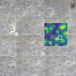
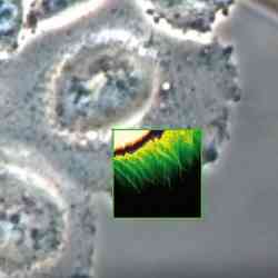
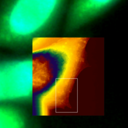
 Living A549 cells - Correlative AFM and STED
 -
 Living Vero cells
 -
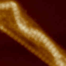
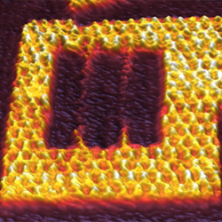 DNA Origami at 150 lines/sec
 -
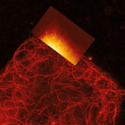
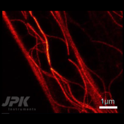 VIDEO - Real-time rapture of microtubules - Correlative AFM and STED measurements
 -
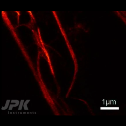 VIDEO - Real-time bending of microtubules - Correlative AFM and STED measurements
 -
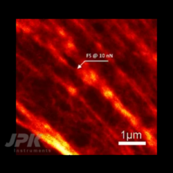 VIDEO - Stimulation of living fibroblast cells - Correlative AFM and STED measurements
 -
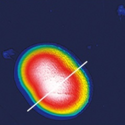
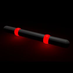 Nanoruler - AFM and STED
 -
 Living A549 cells - Simultaneous AFM and STED
 -
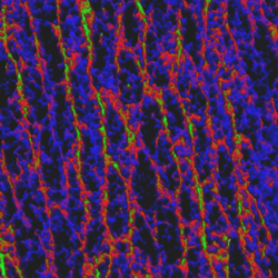
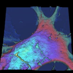 Astrocytes
 -
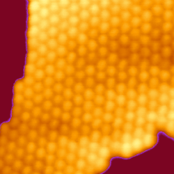
 Imaging of bacteria S-layer with QI™
 -
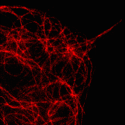
 Simultaneous AFM and STED of Living fibroblasts - Actin Filament Imaging
 -
 Simultaneous AFM and STED of Living fibroblasts - Microtubule Imaging
 -
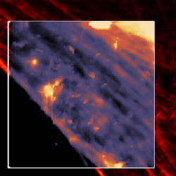
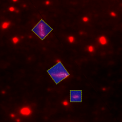
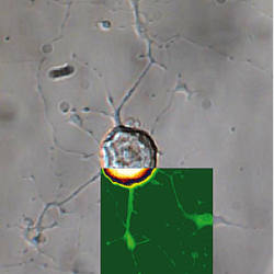
 Cell/particle interaction - AFM with confocal microscopy
 -
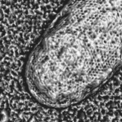
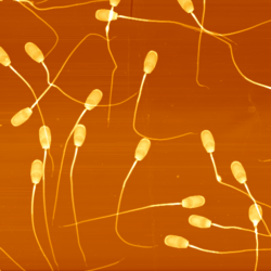 High-resolution imaging on sperm
 -
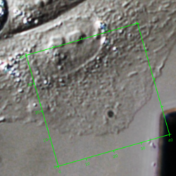 Living CHO cell - AFM with DIC
 -
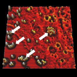
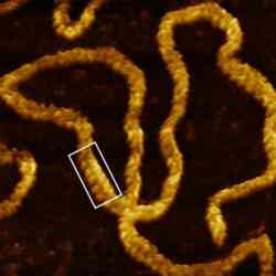
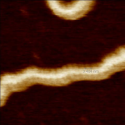
 Rad51 proteins bound to DNA - AFM with fluorescence microscopy
 -
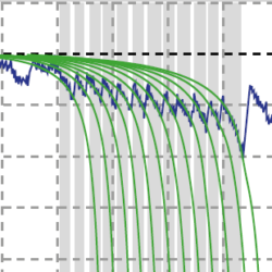 Fibronectin unfolding
 -
 DNA at -25 °C
 -
 CHO cell - AFM with phase contrast
 -
 QI™ DNA - Major and minor grooves
 -
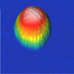
 Recognition microscopy on biotin bead
 -
 Recognition on living keratinocyte cells
 -
 DNA origami - faceman
 -
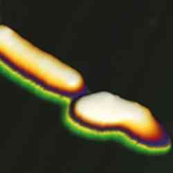
 Twisted insulin fibrils
 -
 Living CHO cells - AFM with fluorescence microscopy
 -
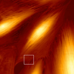
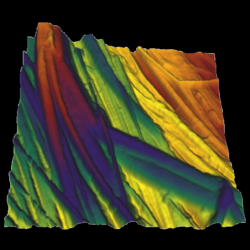 Tendon tissue
 -
 Herpes Simplex Viruses
 -
 Bacteriorhodopsin membrane - QI™ mode
 -
 Cell division E-coli bacteria
 -
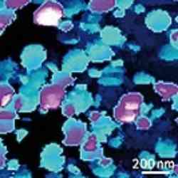
 Human dental enamel
 -
 VIDEO - Live CHO cell dynamics
 -
 Tomato Bushy Stunt Virus
 -
 Plasmid DNA imaged in HyperDrive™ mode in buffer
 -
 KPG7 cell dynamics
 -
 Living Candida albicans - AFM with phase contrast
 -
 HeLa cell in buffer - AFM with STORM
 -
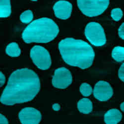
 Melting of lipid domains in buffer
 -
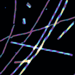
 Collagen in liquid, 70Hz line rate
 -
 Major-minor grooves of λ-phage DNA in liquid
 -
 Bacteriorhodopsin in buffer
 -
 Living fibroblast cell - AFM with phase contrast
 -
 Desulfobulbus bacterial cells
 -
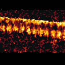
 Bundled collagen fibres
 -
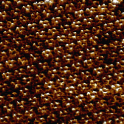
 Pea starch granules
 -
 Herpes Simplex Virus
 -
 Living Cyanobacterium
 -
 Living Escherichia coli bacteria
 -
 Living dorsal root ganglion cells - AFM with DIC
 -
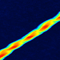
 Twisted amyloid fibrils
 -
 Collagen – AFM with phase contrast
 -
 Glucagon fibre
 -
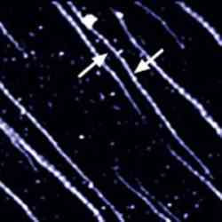
 Fibrillin microfibrils
 -
 Malaria infected red blood cells
 -
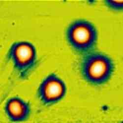
 L929 cell filipodia
 -
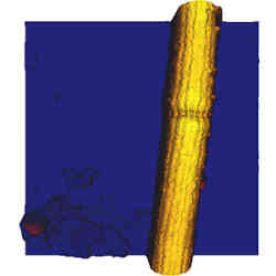
 Single waste water bacterium
 -
 OmpF protein crystal
 -
 Lipid bilayer - AFM with confocal microscopy
 -
 MDCK cells - AFM with confocal microscopy
 -
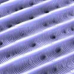
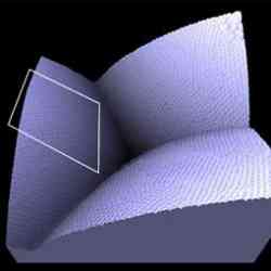 Moth's eye
 -
 Human lymphocyte chromosomes
 -
 Unfixed collagen in buffer
 -
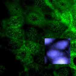
 Ptk2 cells - AFM with fluorescence microscopy
 -
 Living fibroblast cells – AFM with phase contrast and fluorescence
 -
 Living fibroblast cell
 -
 Lipid vesicles
 -
 Moth wing scale
 -
 Collagen manipulation
 -
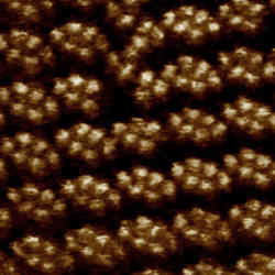
 Gold clusters in water
 -
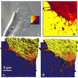
 REF52 cells - AFM with fluorescence microscopy
 -
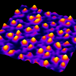
 High-resoution image of HPI layer